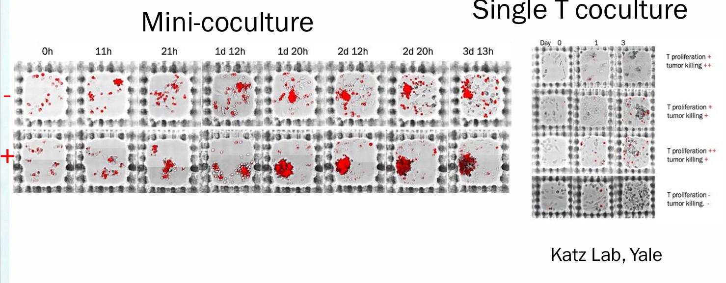
Katz Lab, Yale University
Nanobody MET CAR-T cells show efficacy in solid tumors — January 2026 preprint
Nanobody MET CAR-T cells show efficacy in solid tumors
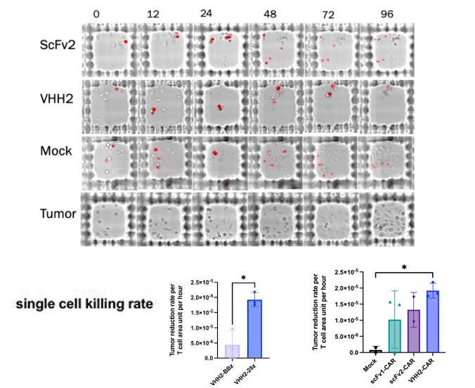
MET overexpression is associated with poor prognosis across many solid tumors. This study tested the efficacy of MET-targeting VHH CAR-T cells as an alternative to conventional scFv-based constructs — which often suffer from tonic signaling and instability in solid tumor environments. VHH-CAR-T cells were evaluated using hydrogel microwell-based cellular kinetics on TROVO, tracking real-time cytotoxicity and killing behavior across thousands of individual co-cultures.
Read the Full Article (PMC) →
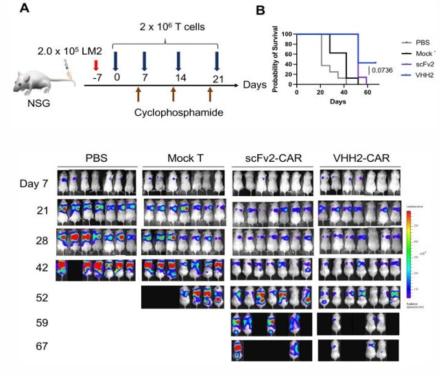
In vivo tumor control — VHH2-CAR vs. scFv constructs in NSG mouse TNBC model

Per-clone killing kinetics across CAR-T constructs in microwell co-culture
Key Findings
Intermediate avidity clones outperformed high-avidity ones in vitro — only discoverable through per-clone kinetic tracking, not bulk assays.
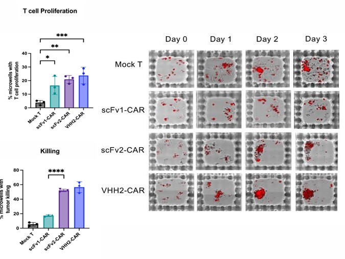
VHH-CAR-Ts showed superior biochemical stability and favorable cytokine profiles vs. traditional scFv-based designs.
TROVO microwell co-cultures enabled direct measurement of cytotoxicity and T cell behavior in real time across thousands of individual interactions.
Potent and prolonged tumor growth control in metastatic triple-negative breast cancer models, both in vitro and in vivo.
TROVO in Action
Real workflows, real data — generated using TROVO in active research programs.
TIL Potency Assessment — Distinguishing Responders from Non-Responders
Katz Lab, Yale University
Identifying which patient TILs are capable of sustained tumor killing is one of the hardest problems in clinical immunotherapy. Surface markers alone don't answer this question. TROVO seeds patient TILs and tumor cells into thousands of microwells, tracks killing behavior longitudinally, and compares each co-culture against 50 matched tumor-only controls per microwell. The result is a statistically rigorous Z-score for each clone — derived from the patient's own sample, with no predefined markers required.
In data from the Katz Lab at Yale, TROVO successfully distinguished TILs from responding versus non-responding patients using as few as ~10,000 cells — a cell input level incompatible with traditional bulk functional assays.
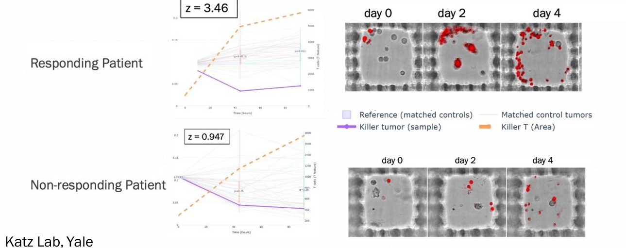
Killer clone abundance (Z-score >2) — responding vs. non-responding patient TILs, two biological replicates
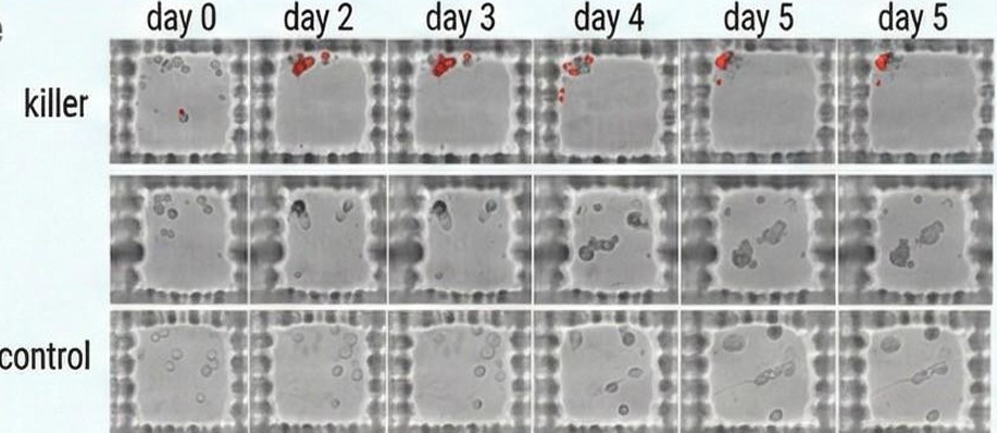
Killer and control microwell trajectories over 5 days — ranked by Z-score
- Native internal controls: Every microwell compared against 50 matched tumor-only control wells — no external reference needed.
- Statistically significant from ~10,000 cells: Rigorous potency metrics from clinical samples too scarce for traditional methods.
- Responder/non-responder discrimination: Killer clone abundance correlates with clinical response in Yale patient cohort data.
- Compatible with downstream characterization: Identified killer populations retrieved intact for NGS, expansion, or re-challenge.
Full TIL Discovery Workflow — From Tissue to Functional Clone Recovery
TROVO supports the complete upstream TIL workflow: tissue dissociation, microwell seeding, longitudinal co-culture imaging, functional clone selection, and live cell recovery — all on a standard 6-well plate, without microfluidics or predefined markers.
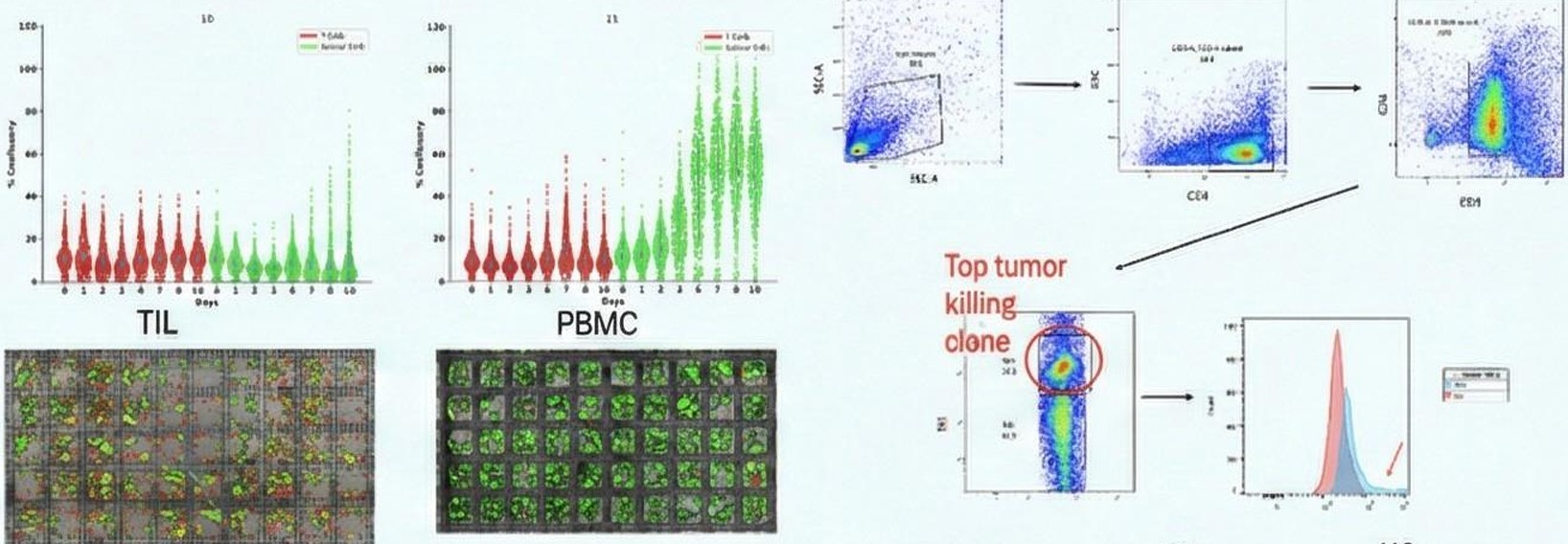
Phenotyping screening and enrichment — TIL vs. PBMC killing index comparison
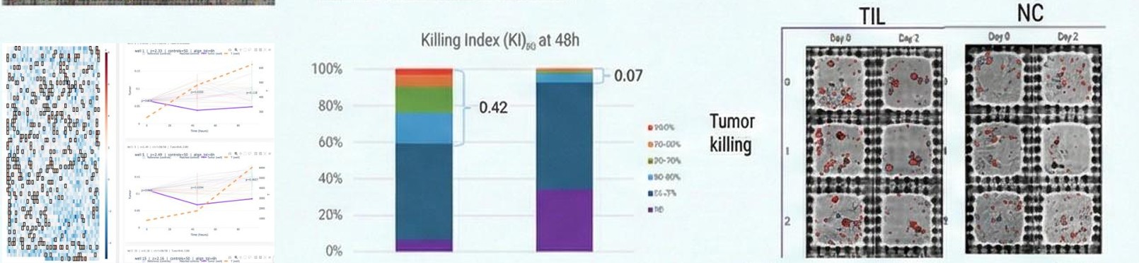
Killing Index (KI₅₀) at 48h — TIL vs. normal control, with tumor killing progression over days
Workflow Steps
- Dissociate tumor tissue to obtain TIL and tumor cell suspension
- Seed cells into PEGDA hydrogel microwells — single cell encapsulation per well
- Image longitudinally over 2–5 days using TROVO fluorescence and brightfield imaging
- Rank microwells by Z-score killing index against 50 matched tumor-only controls
- Select high-performing microwells for light-induced capture gel polymerization
- Wash away uncaptured cells; enzymatically dissolve gel to release viable recovered cells
- Proceed to NGS, expansion, or downstream re-challenge assays
Clone ranking by Z-score — killers vs. controls over time
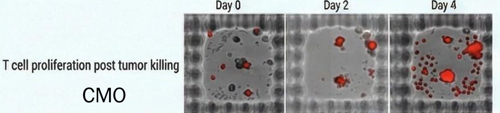
T cell proliferation post tumor killing — Day 0 through Day 4
- CMO-compatible workflow: Designed for clinical manufacturing contexts where sample scarcity and process reproducibility are critical.
- Function-first selection: Cells selected based on observed killing behavior — not surface marker assumptions.
- Intact transcriptome on recovery: Fluidics-free capture means no shear stress — RNA integrity preserved for downstream sequencing.
CAR-T and TCR-T Clone Selection Based on Tumor Co-Cultures
Standard activation markers don't reliably predict which CAR-T or TCR-T constructs will sustain killing over time. TROVO tracks cytotoxicity and persistence kinetics live over 12 days — with tumor rechallenge every 3 days — to separate true persistence clones from short-term burst killers before any downstream investment.
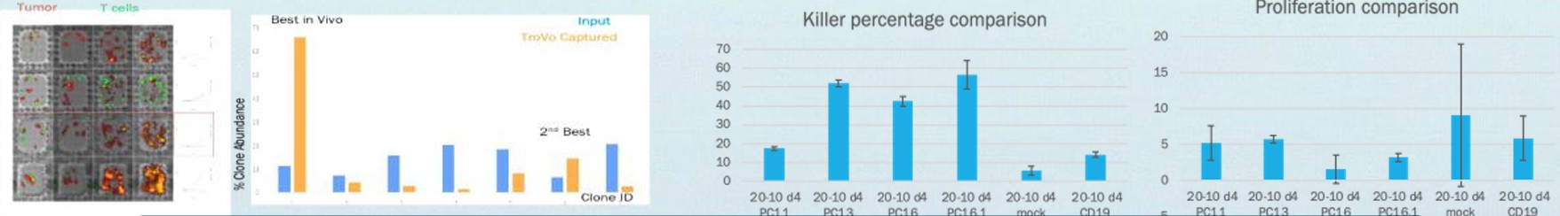
Killer percentage and proliferation comparison across CAR-T constructs
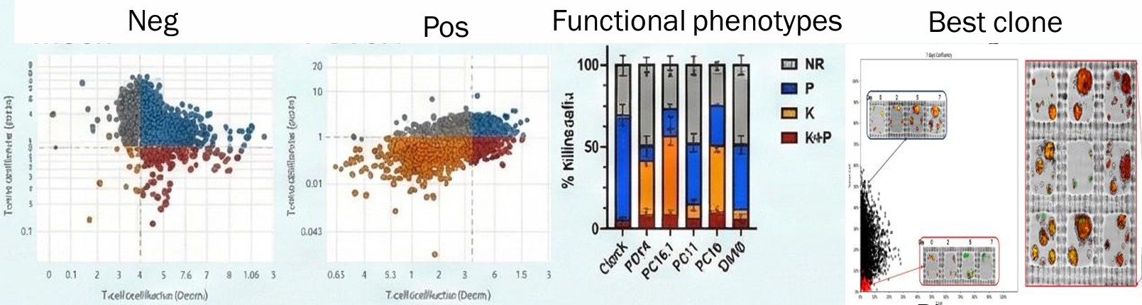
Functional phenotype profiling — NR, P, K, K+P populations across constructs
- 12-day persistence assay: Track the same clones across multiple tumor rechallenge cycles — identify serial killers vs. early exhausters.
- 12,000–18,000 microwells per 6-well plate: Simultaneously monitor thousands of individual co-cultures without sacrificing resolution.
- Functional Z-score ranking: Objective, quantitative clone comparison based on direct behavioral data — not proxy markers.
- Light-induced capture: Retrieve specific viable clones directly for downstream NGS or expansion.

T cell (red) and tumor cell (green) co-culture — 12-day time course on hydrogel microwells

Capture gel workflow — selected microwells encapsulated, unwanted cells washed, viable cells released
T cells recovered from 12-day co-cultures maintained normal phenotypic expression and full functionality post-expansion — confirming that fluidics-free capture does not compromise cell health or downstream performance. This work was supported by NIAID R44AI147734 and NCI 75N91022C00061.
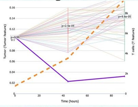
Predator-Prey Kinetics — Intrinsic and Cooperative Tumor Killing
TROVO's per-clone kinetic data can be fit to Lotka-Volterra predator-prey equations to derive a quantitative killing rate constant (keff) for each individual clone. This separates intrinsic single-cell killing capacity from cooperative "wolf-pack" killing at higher effector densities — and the resulting in vitro kinetic profiles correlate directly with in vivo tumor control and survival outcomes.
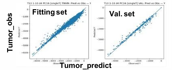
Lotka-Volterra model — tumor observed vs. predicted across 3,000 microcultures, R² >0.95
Single-cell killing kinetics — intrinsic k_eff at E:T 1:10, differentiating constructs independent of cooperative effects
Single T co-culture — four distinct killing phenotypes
Collaborative killing at E:T 20:10 — population-level clearance and proliferation
In vivo validation — k_eff profiles correlate with NSG mouse survival outcomes
- 3,000 microcultures, 144-hour assay: R² >0.95 on held-out validation set — model fit across both fitting and validation sets.
- Intrinsic vs. cooperative killing separated: E:T 1:10 isolates individual clone capacity; E:T 20:10 reveals population dynamics.
- In vivo correlation: In vitro keff predicts in vivo tumor control and survival in NSG mouse models.
High-Throughput B Cell Screening for Native Protein Binding
Single-readout B cell screens are prone to false positives from sticky targets or non-specific IgGs. TROVO enables a two-round multi-signal screening process — first against target peptide on beads, then against full-length native protein on cells — to identify "double-positive" clones that bind in both conformations before committing to sequencing.

PBMCs co-incubated with target beads, anti-IgG secondary staining, positive microwell selection and capture

Two-round screening — double-positive clones binding both target peptide and full-length native protein
- ~0.04% positive hit rate detected: Demonstrated sensitivity to rare secretors in primary PBMC repertoires across multiple seeding densities.
- Secretor detection in under 2 hours: From seeding PBMCs to identifying positive microwells in a single session.
- Native conformation specificity: Two-round screening ensures antibodies bind full-length protein in its native form — not just synthetic peptides.
- No microfluidics: Same resolution as nanopen-based platforms without the complexity, cost, or cell stress.
Screening Protocol
- Immunize animals and obtain PBMCs
- Coat streptavidin beads with biotinylated target peptide
- Seed PBMCs and beads into microwells; pre-stain PBMCs with green fluorescence; incubate 2 hours
- Remove excess media; add anti-rabbit IgG Alexa647 for staining; image with TROVO
- Select microwells with high red fluorescent signal (positive secretors)
- Add liquid capture gel; photo-crosslink in selected microwells; wash away uncaptured cells
- Dissolve capture gel enzymatically; retrieve cells for RT-PCR, Sanger sequencing, or expansion
Live Cell Enrichment from Tissue for Single-Cell RNA Sequencing
Preparing clinical tissue samples for single-cell sequencing is a bottleneck — most methods require large tissue input, risk clogging from debris, or damage fragile cells through high-pressure sorting. TROVO's image-guided live cell enrichment is designed for exactly the cases where standard bulk approaches fail.

Workflow: fresh or frozen tissue → digestion → microwell seeding → dead cell capture → live cell retrieval

% fraction reads in cells — TROVO-enriched vs. unenriched control across breast, lung, and colon tissues
- 4 hours for 3 samples: From raw tissue to single live cell suspension — batch processing compatible.
- Requires only 10⁴ cells: Designed for 5mm tissue pieces and rare clinical biopsies where input is limited.
- Fluidics-free at 4°C: No clogging, no shear stress, intact transcriptome — optimized for fragile edited lines and primary tissue.
- Proven data quality improvement: Higher % fraction reads in cells vs. unenriched controls across multiple tissue types in 10X Genomics scRNA-seq.
- Tissue-specific features preserved: Cell type clustering and tissue-specific distributions maintained in downstream analysis.
On-Demand Recordings
Full-length webinars featuring TROVO collaborators presenting their research.
Integrated High-Throughput Functional Profiling of Myeloid Cells in Pancreatic Cancer
Dr. Won Jin Ho, Johns Hopkins University
Dr. Won Jin Ho presents the DEFINE workflow — linking real-time functional killing kinetics directly to high-plex Imaging Mass Cytometry (IMC) protein data. By performing in situ proteomic profiling of rare clones within hydrogel microwells, DEFINE answers a question flow cytometry cannot ask: not just what a cell is, but what it did. This session covers myeloid heterogeneity across PDAC tumor sites, how specific myeloid subsets suppress or enhance T cell-mediated killing, and how TROVO enables the functional co-culture assays that make this indexing possible.
Functional Selection of CAR-T Clones from 2D and 3D Brain Tumor Co-Cultures
Dr. Xiujian Ma, City of Hope
Scientists at City of Hope demonstrate how TROVO supports CAR-T clone selection in both 2D and 3D brain tumor co-culture models, including glioblastoma. This session covers the challenge of CAR-T clone selection for solid tumors, how TROVO's real-time cytotoxicity imaging enables functional identification without predefined biomarkers, and how AI-driven kinetic analysis makes the approach scalable for CAR-T development programs.