Turn your 6-well plate into thousands of individual experiments.
TROVO uses light-based hydrogel lithography to print customizable microwells directly onto standard cell culture plates — no new labware, no proprietary consumables beyond the gel reagent. The entire plate prints in under an hour, and the resulting microwell array sits in your incubator like any other plate.
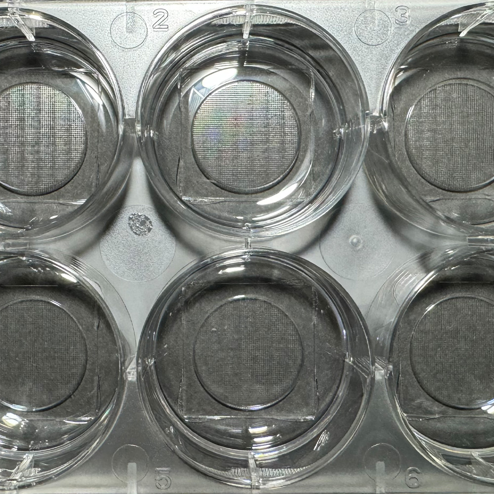

150×100µm hydrogel microwells printed on a standard glass-bottom 6-well plate
Print Specifications
Configurable microwell sizes
- Standard labware: Works on 1-, 6-, 24-, and 96-well plates, chamber slides, and glass slides — no proprietary plate formats required.
- Any size or shape: Microwell geometry is configurable to your experimental needs — round, square, or custom footprint.
- Biocompatible PEGDA hydrogel: The microwell walls are inert, non-toxic, and compatible with standard cell culture reagents and matrices.
- Culture medium is shared: At 200µm wall height, medium, oxygen, and cytokines are freely accessible across the array — maintaining physiological conditions throughout the assay.
Discover
Watch what your cells actually do — over hours, days, or weeks.
Once cells are seeded, TROVO images the entire array automatically at defined timepoints, capturing fluorescence and brightfield data per microwell. Every clone has its own time-course record. You're not averaging a population — you're watching individual co-cultures in parallel, measuring cytotoxicity, persistence, proliferation, exhaustion, and secretion simultaneously.
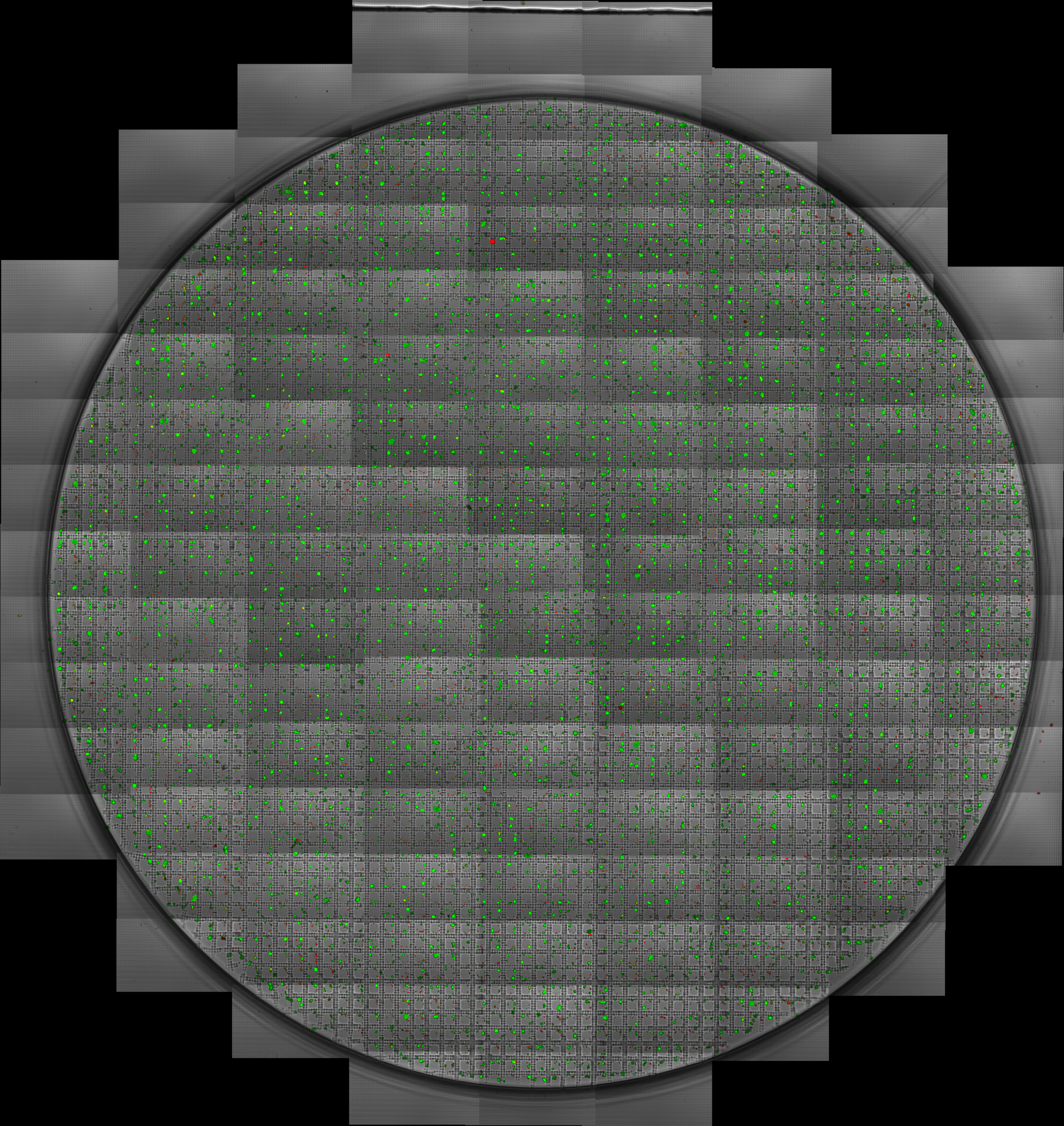
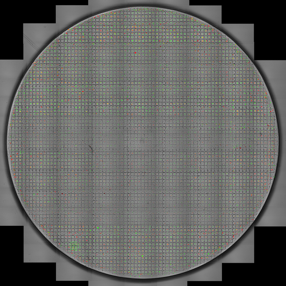
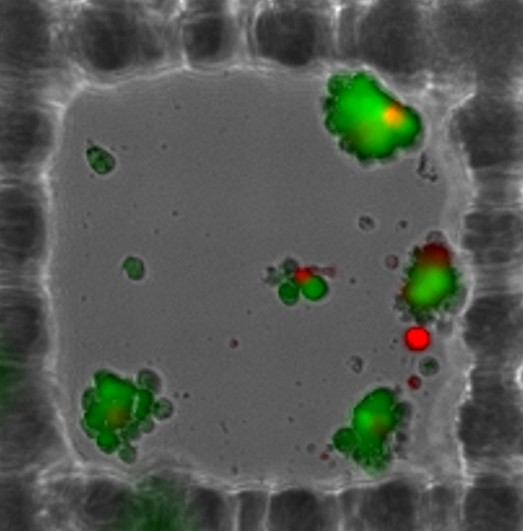
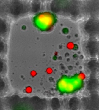
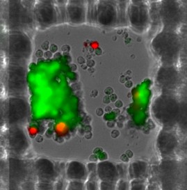
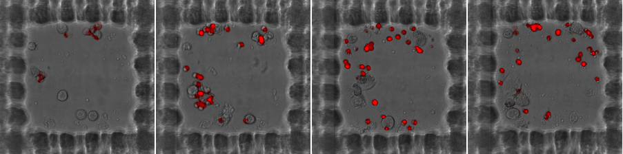
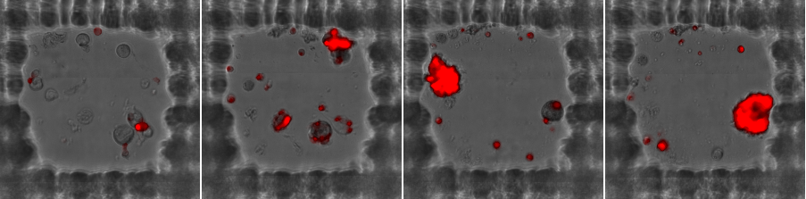
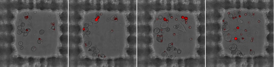
A full well of co-culture microwells, imaged in fluorescence
Zoomed in — the array
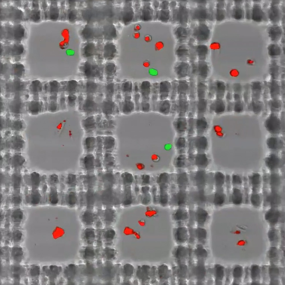
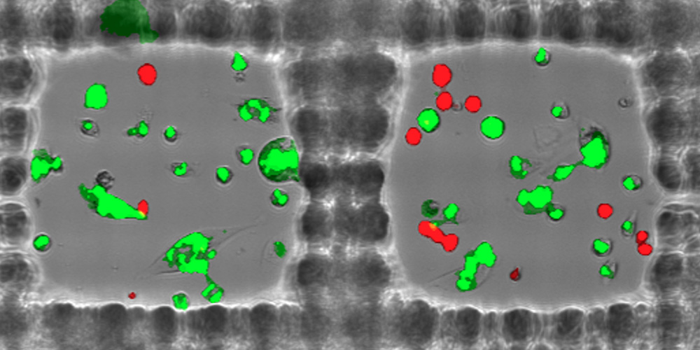
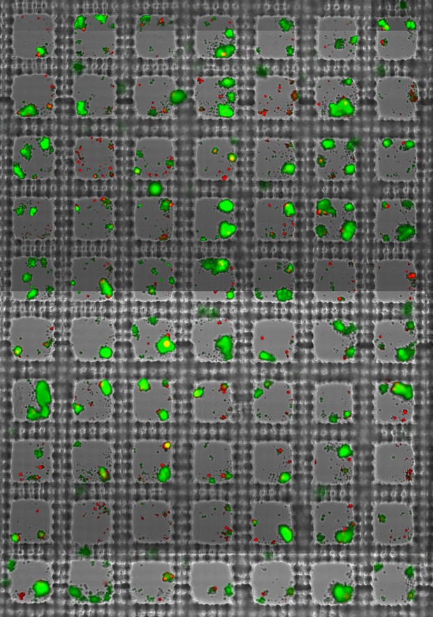
Zoomed in — individual microwells
Zoomed in — individual microwells
Zoomed in — individual microwells
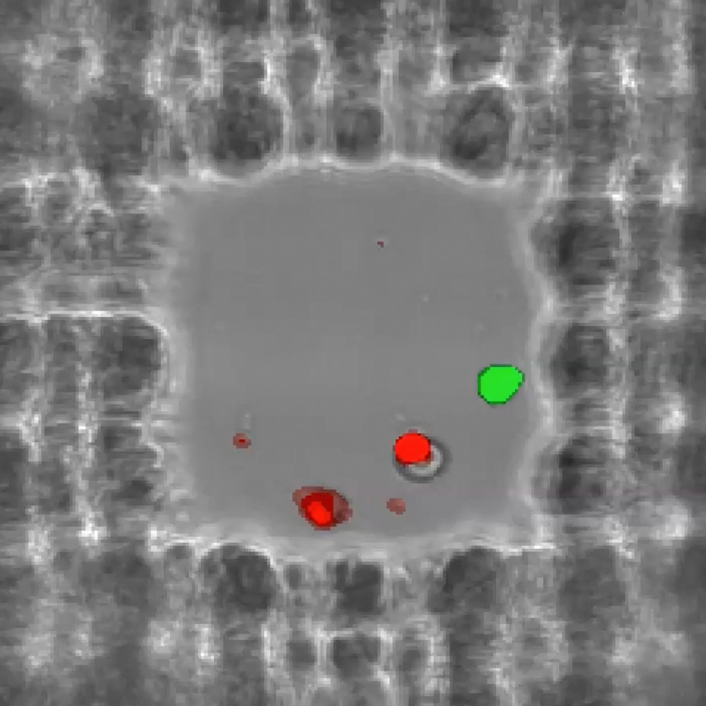
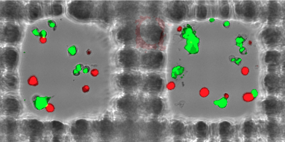
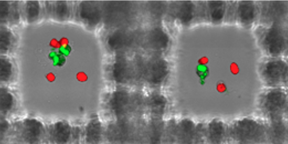
Killer vs. Non-Killer Wells
The same imaging works whether your cells are adherent or in suspension — TROVO doesn't require a different setup for either. What matters is what shows up in the data: microwells where the effector cells killed their targets, and microwells where they didn't.
Imaging Specifications
What TROVO measures
- Cytotoxicity: Per-microwell tumor cell killing over time
- Persistence: Which clones keep killing vs. exhaust after one hit
- Proliferation: T cell expansion concurrent with killing
- Exhaustion: Longitudinal decline in killing activity
- Secretion: Fluorescent signal from secreted antibodies or cytokines
- Migration: Cell movement within and across microwell boundaries
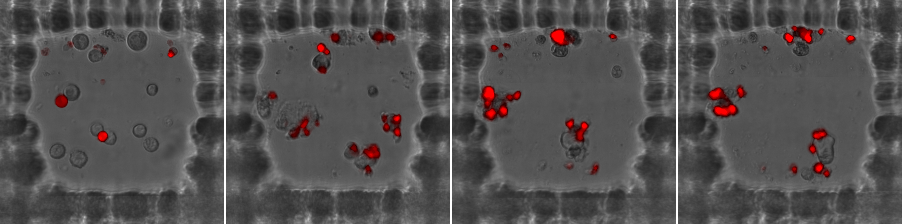
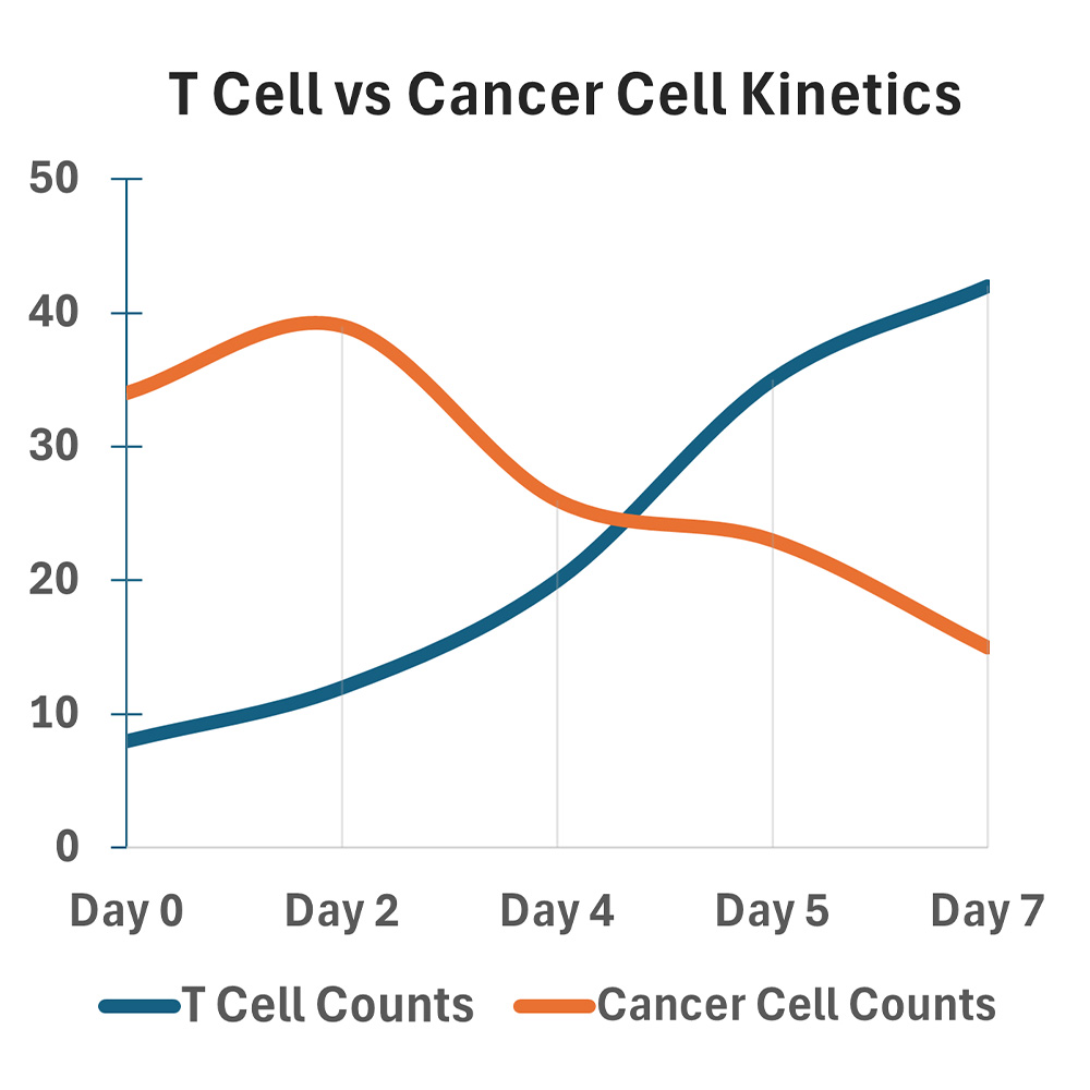
Why longitudinal tracking matters
Flow cytometry gives you a snapshot. Incucyte gives you a population average. Neither tells you which specific clone in your sample showed 12-day persistence — or which one looked great on Day 3 and exhausted by Day 7.
TROVO tracks the same microwells across the full assay duration, generating a complete behavioral history for every clone. That's the data you need to rank them meaningfully and select the right ones for recovery.
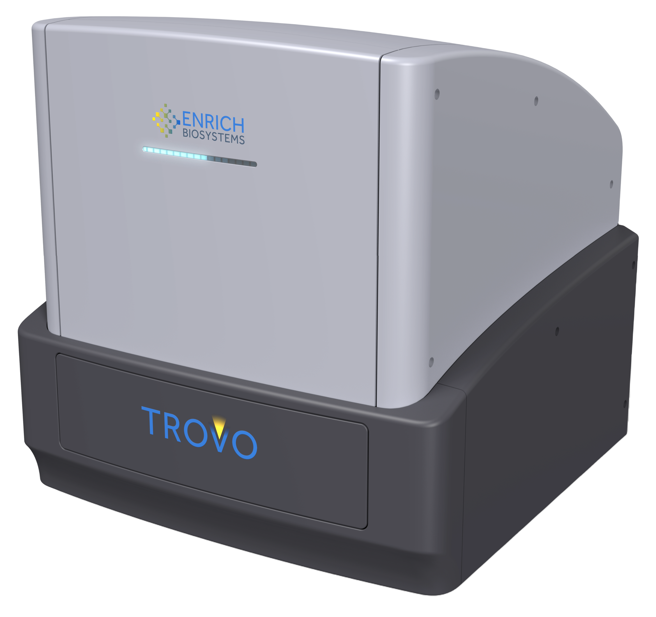
The TROVO system — 18"×18"×18", 55 lbs, designed to sit in a standard cell culture incubator
Poisson distribution generates its own controls
When cells are seeded at appropriate density, Poisson statistics naturally populate the array with T-cell-only wells, tumor-only wells, and a gradient of effector-to-target ratios — all side by side on the same plate, imaged under identical conditions.
This eliminates plate-to-plate variability and gives you the statistical power of a 50-plate experiment at a fraction of the cell input. Every microwell in the culture is compared against 50 matched tumor-only control wells, and Z-score >2 represents statistically significant tumor killing.
Retrieve
Recover the cells that earned it — alive, intact, ready for what's next.
Once the imaging phase identifies microwells of interest, TROVO's light-induced capture gel selectively encapsulates those populations in place. Unwanted cells are washed away. The capture gel is dissolved enzymatically to release the recovered cells — which arrive viable, with intact transcriptome, and ready for NGS, expansion, or re-challenge. No FACS. No microfluidics. No shear stress.
Capture gel encapsulation
How the capture works
A liquid biocompatible capture gel is applied across the plate surface. TROVO then projects a 405nm laser through an LCD screen beneath the plate, polymerizing the gel only in the selected microwells — leaving every other microwell unaffected.
The plate is then washed, removing all cells outside the capture gel. The solidified gel containing the target populations is enzymatically dissolved, releasing the cells into solution for downstream use.
- Selection is behavioral, not marker-based: You're recovering what you watched perform — not guessing based on surface phenotype.
- No physical stress: Cells never experience high-pressure fluidics or droplet generation — transcriptome integrity is preserved.
- Flexible downstream: Recovered populations go directly to NGS, REP expansion, re-challenge assays, or direct-to-PCR workflows.
From imaging to selection — the analysis pipeline
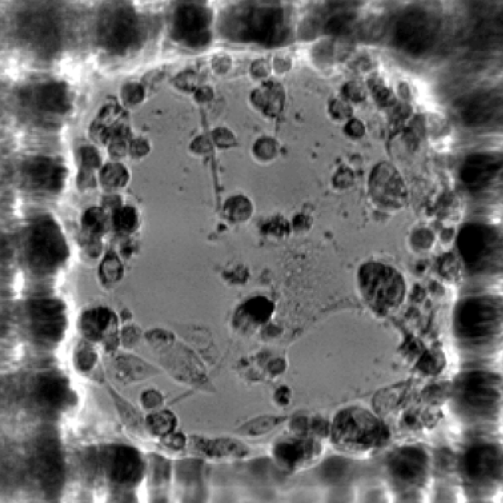
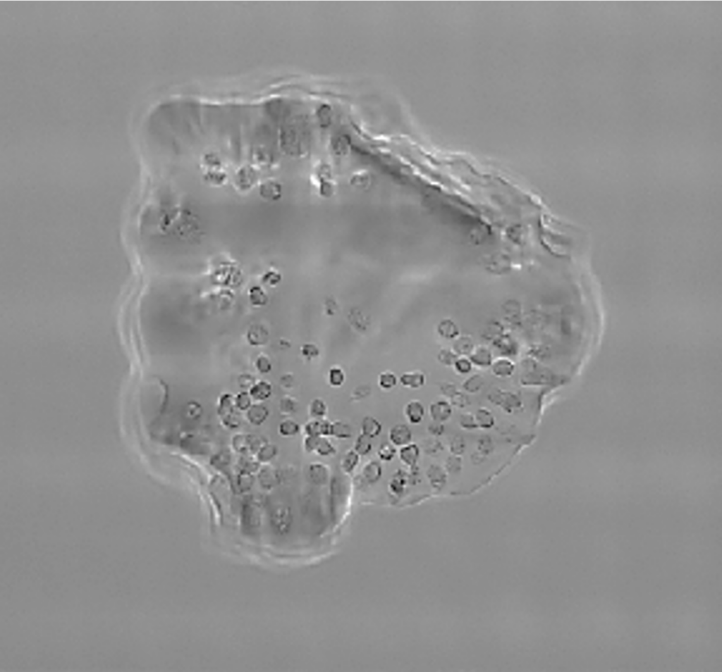
Brightfield imaging of cells growing in microwells over time
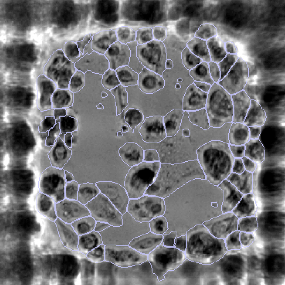
Cell boundaries segmented using deep learning (CellPose3)
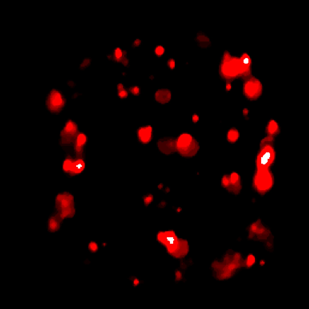
Fluorescent channels distinguish T cells from tumor cells in co-culture
Automated analysis extracts per-clone potency, proliferation, and killing criteria
Top microwells selected and encapsulated in protective gel for retrieval
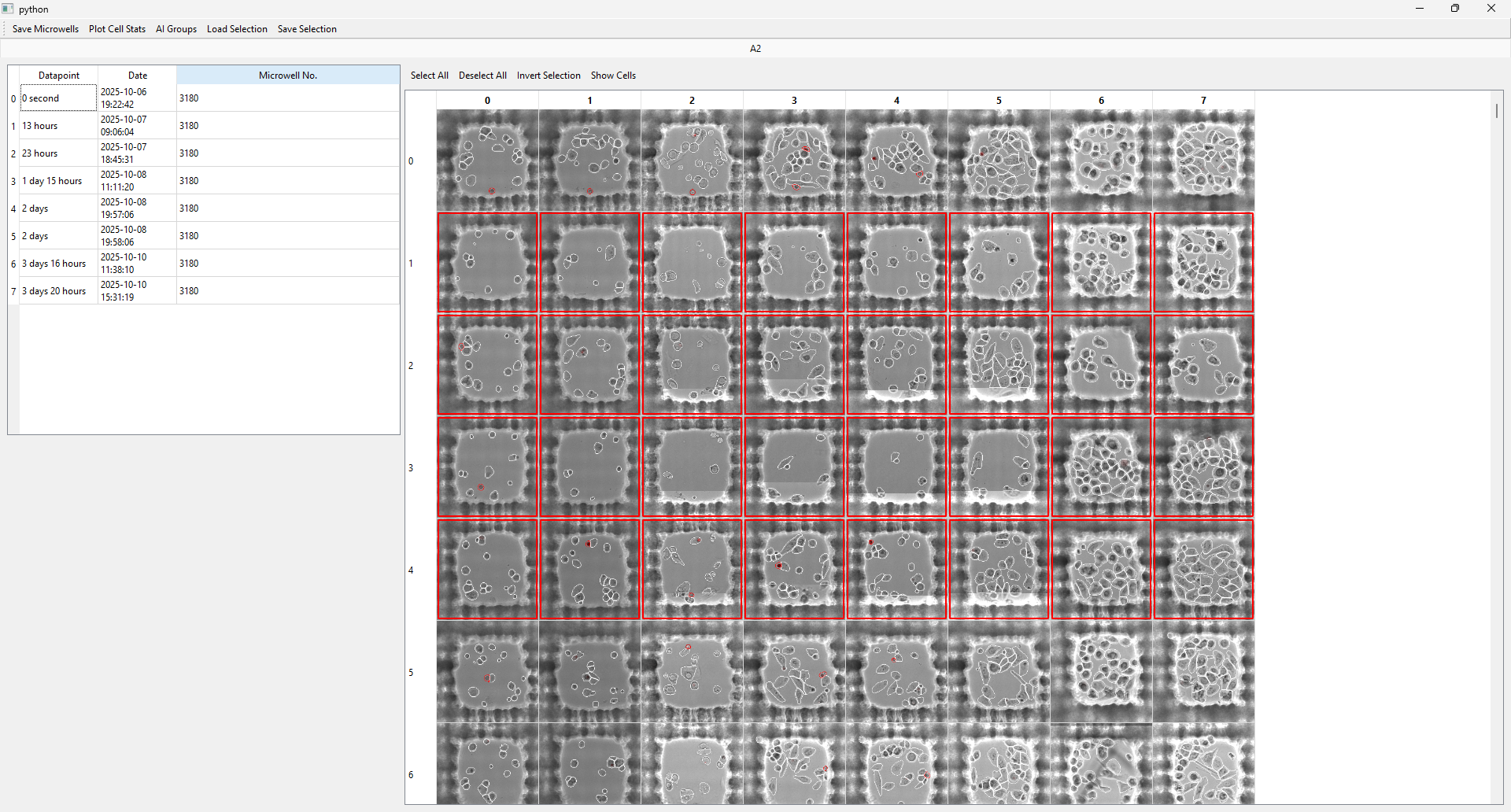
TROVO's software — review per-microwell data and select cells for retrieval
Retrieval at a glance
See the full workflow in your context.
Talk to our team about your specific application — we'll walk through how Print, Discover, and Retrieve map to your actual experiment.